When imaging detects a region of interest or suspicion, it can also be used to direct selective biopsies to obtain very small tissue samples for further laboratory analysis (pathology). The use of imaging together with pathology gives the most accurate information about the size, location and aggressiveness of any cancer thus identified.

Breast Cancer
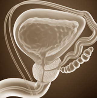
Prostate Cancer
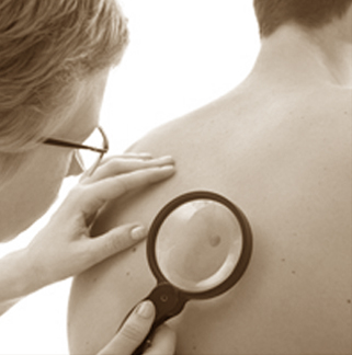
Skin Cancer

Thyroid
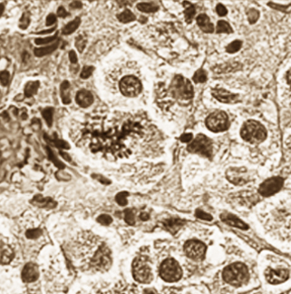
Melanoma
Breast Cancer
The current standard in screening for breast cancer is mammography. However, this imaging tool misses some breast tumors, especially in women with dense breasts. Published data suggests that sonography can play an important role in detecting tumors that mammography misses. In fact, over 94% of cancers seen only on ultrasound were invasive tumors with average size of 10 mm, and in the studies where staging was detailed, 91% were node negative, meaning it had not spread and complete cure was possible due to early detection.
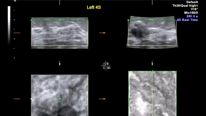
Power Doppler Sonography, such as that used by Dr. Bard, adds increased accuracy to breast imaging evaluation over ordinary ultrasound because it shows higher blood flow speeds, often a sign of cancerous activity in the breast. Studies have shown that suspicious blood flow identified by pre-surgery Power Doppler scans corresponds very well with the size, location and aggression of actual tumors that are then surgically removed. Thus, Power Doppler brings an important clinical dimension to breast cancer detection. 3D sonography clearly shows tumor margins and 3D Doppler affords an index of cancer aggression and metastatic potential.
While it has not been established that sonography should be used in place of mammography, ultrasound offers the following advantages:
Click MORE for complete info on BREAST CANCER
- Highest accuracy in dense (lumpy, cystic) breasts
- Non-invasive-no radiation exposure
- Cost effective
- Can distinguish cysts (fluid-filled masses) from cancerous tumors without needle sampling
- Ease of image guidance for breast biopsy
If there is a down side, it is the shortage of medical personnel skilled at reading breast sonograms. This suggests the importance of seeking expert sonographers when breast ultrasound is indicated or desirable as a complement to mammography.
It has not been determined if there are specific groups of women who would most benefit from ultrasound breast exams alone, or in conjunction with mammogram screening. Women who should consider ultrasound scanning of the breast include those at risk of breast cancer because of personal or family history and the presence of fibrocystic (dense) breast tissue which increases cancer risk by 400%. Dr. Bard’s practice is accredited by the American College of Radiology for breast imaging including mammography.
Did you know…

Breast feeding mothers with suspect symptoms can be painlessly checked for tumors with ultrasound screening. Infected pockets can be drained under ultrasound guidance reducing need for antibiotics.
 BARD CANCER DIAGNOSTICS is founded on the commitment to explore and implement the latest diagnostic technologies as a means of building the proper treatment strategy of many types of cancers. We also specialize in the PREVENTION solutions for our patients who strive to maintain a health-conscious lifestyle as well as those who are at increased risk of certain diseases by confirming that their efforts to prevent disease are working.
BARD CANCER DIAGNOSTICS
121 E. 60th St. Suite #6A New York, NY 10022
212.355.7017
Info@bardcancercenter.com